The cells lie apex-to-apex with cells of the pigment layer that extend to the margin of the optic cup and cover the developing ciliary region and iris. The ciliary body arises from neuroectoderm of the anterior optic cup and its associated mesoderm. Neuroectoderm forms the outer pigmented and inner nonpigmented epithelia that cover the ciliary body and ciliary processes; mesenchyme forms the ciliary muscle, stroma, and blood vessels. As the ciliary muscle differentiates, folds appear in the outer layer of neuroectoderm, external to the margin of the optic cup. Each fold gives rise to a ciliary process, complete with a core of mesenchyme and blood vessels and covered by two epithelial layers in which the cells are arranged apex-to-apex. Zonule fibers develop within the vitreous and extend from the inner surface of the ciliary process to the lens capsule. The double layer of epithelium covering the iris develops from the double cell layer of the optic cup. The smooth muscle of the sphincter and dilator muscles of the iris is unusual in that it develops from neuroectoderm. An ingrowth of mesenchyme at the edge of the optic cup provides the stroma and vasculature of the iris. The optic nerve develops from axons of retinal ganglion cells, neuroectodermal cells, and mesoderm associated with the optic stalk. The mesoderm gives rise to the vascular components and connective tissue of the nerve, including the meninges. At first, the optic stalk contains an open ventral groove, the choroid fissure, through which mesenchyme enters the optic cup. Hyaloid vessels that supply the lens and inner surface of the retina during their development and axons from the brain to the retina also pass within the groove. As the choroid fissure closes, the optic stalk is transformed into the optic nerve with the central retina (the previous hyaloid) artery at its center. That part of the hyaloid artery that supplied the lens degenerates and is resorbed. The mesenchyme filling the posterior aspect of the optic cup becomes the gelatinous, transparent vitreous. It is strengthened by the mesenchymal component that gives rise to fibroblasts and collagenous septae that pass among the neuroglial cells. Accessory Structures Folds of integument adjacent to the eyeball differentiate to form the eyelid. Ectoderm on the exterior of the developing eyelid becomes the outer layer of stratified squamous epithelium (epidermis): that covering the cornea and anterior part of the sclera forms the conjunctiva. Eyelashes and sebaceous, sweat, and tarsal glands develop along the edge of each eyelid while they still are fused. The follicles of the lashes and their associated glands arise as epidermal ingrowths that then develop as hairs elsewhere in the body. The lacrimal sac and nasolacrimal duct first appear as a solid outgrowth Table 20-1. Embryonic Layer Neuroectoderm Surface ectoderm Mesenchyme Adult structures of epithelium from the nasolacrimal groove; a second growth from the epithelium of each eyelid joins it. The distal end of the cord grows toward the nasal cavity and fuses with the nasal epithelium prior to acquiring a lumen. Neural retina, pigment epithelium, epithelium of iris, dilator and sphincter pupillary muscles, nervous and neuroglial elements of optic nerve Epithelium of cornea, lens Substantia propria, endothelium of cornea, sclera, choroid, stroma and vessels of iris, ciliary body, ciliary processes, ciliary muscle, sheaths of optic nerve; anterior, posterior, and vitreous chambers Summary the corneoscleral coat, together with the intraocular pressure of the fluid contents within the eye, maintains the proper shape and size of the eyeball. Light entering the eye must cross several transparent media (cornea, aqueous humor, lens, and vitreous body) before reaching receptors in the retina. There are no blood vessels in the transparent elements, which rely on diffusion of materials for their nutrition. Peripheral regions of the cornea receive nutrients from adjacent vessels in the limbus; the remainder of the cornea depends on diffusion of nutrients from the aqueous humor.
Occasionally the gap is filled by formation of hyaline cartilage, which then undergoes endochondral ossification to achieve cortical union. More frequently, the bone that initially unites the broken ends is a network of woven bone formed by intramembranous bone formation. The last act in repair is the resorption of excess bone and the remodeling of newly formed and dead bone that is replaced by lamellar bone. Bone morphogenic proteins act at all the important steps in the cascade of events that form new bone: chemotaxis of progenitor cells, mitosis, differentiation and proliferation of chondrocytes and osteoblasts, stimulation of extracellular matrix formation and binding to specific matrix molecules. Summary Cartilage serves as a rigid yet lightweight and flexible supporting tissue. It forms the framework for the respiratory passages to prevent their collapse, provides smooth "bearings" at joints, and forms a cushion between the vertebrae, acting as a shock absorber for the spine. Cartilage is important in determining the size and shape of bones and provides the growing areas in many bones. Its capacity for rapid growth while maintaining stiffness makes cartilage suitable for the embryonic skeleton. About 75% of the water in cartilage is bound to proteoglycans, and these compounds are important in the transport of fluids, electrolytes, and nutrients throughout the cartilage matrix. It provides attachment for muscles of locomotion, carries the joints, serves as a covering to protect vital organs, and houses the hemopoietic tissue. Osteocytes are the dominant cells of mature bones and are responsible for maintaining the matrix. They also aid in regulating the calcium and phosphorus levels of the body and play a role in the resorption of bone. They destroy the ground substance and collagen and release minerals from the matrix. These cells elaborate lysosomal enzymes and contain high concentrations of citrate, which is involved in mobilizing calcium from bone. The initial stage of bone resorption by osteoclasts is extracellular: glycosaminoglycans of the matrix are degraded, permitting fragmentation of the bone. The ruffled border of the osteoclasts increases the surface area and seals off the area of resorption and allows a local environment conducive to the digestion of bone. Longitudinal growth of a developing bone depends on the interstitial growth of cartilage in the zone of proliferation of the epiphyseal plate and on the enlargement of chondrocytes in the zone of maturation and hypertrophy nearby. Cartilage of the epiphyseal plate is under the influence of growth hormone secreted by cells in the anterior pituitary gland. Thyroid hormone modulates the activity of growth hormone, and at puberty, androgens and estrogens contribute to masculinization or feminization of the skeleton, respectively. Increased width of the bone is achieved by appositional growth and the deposition of bone at the boney collar. The first primitive trabeculae observed during endochondral bone formation consist of cores of cartilage covered by bone. They are formed in the region where the boney collar is thinnest and serve as internal struts to support the developing bone until the collar is thick enough to provide support. As the collar increases in thickness, osteoclasts remove bone at the endosteal surface, thereby enlarging the marrow cavity. Although most joints serve to allow movement, sutures are immobile and act only to unite bones. Sutures in the young provide areas where bone can grow, and it is only after they have been replaced by boney unions that their uniting functions become paramount. Even the articular cartilages of synovial joints serve as areas of growth for subchondral bone during development. The cartilaginous joints of the spine, although permitting some degree of motion, serve primarily as shock absorbers to dampen and distribute mechanical forces acting on the spine. This is mainly the function of the nucleus pulposus, which, because of its high water content, can act as a water cushion. The fibrous capsule and ligaments provide a tough, unyielding binding for the articulating bones yet remain flexible enough to allow movement of the joint. The synovial membrane mainly produces the lubricant of the joint, the synovial fluid, but also may participate in removing particulate matter from the joint cavity. Synovial villi increase the surface area for secretion and absorption, while the larger folds serve as flexible pads that can accommodate to the changing shape and size of the joint space and its recesses during movement.
The parietal layer of the epicardium consists of connective tissue lined by mesothelial cells that face those covering the visceral epicardium. The two epithelial lined layers are separated only by a thin film of fluid, produced by the mesothelial cells, that allows the layers to slide over each other during contraction and relaxation of the heart. Each cardiac cycle is initiated by the spontaneous generation of an action potential by cardiac muscle cells forming the sinoatrial node. The sinoatrial node is located in the epicardium at the junction of the superior vena cava and right atrium and forms an ellipsoid strip about 13 mm long and 3 mm wide. Nodal cells are smaller than ordinary cardiac muscle cells and contain fewer and more poorly organized myofibrils. Because of the high sodium ion concentration in the surrounding extracellular fluid, these ions normally tend to leak into the sinus muscle fibers. It is the inherent leakiness of the plasmalemma of sinus nodal fibers to sodium ions through special sodium ion channels (If channels) that is related to the self-excitation phenomenon. As a result of this leakage, the resting membrane potential of the sinus nodal fibers is lower (-55 to -60 millivolts) in comparison with normal cardiac myocytes of the ventricles (-85 to 90 millivolts). As a result of the less negativity of the resting potential, the fast sodium channels are generally inactive and only the slow calcium-sodium channels open resulting in the development of an action potential. The ends of the cardiac muscle fibers constituting the sinoatrial node are linked directly to the adjacent ordinary atrial cardiac muscle cells. The spontaneously generated action potential initiated in the node cells then spreads throughout the entire atrial muscle mass to the atrioventricular node located in the posterior wall of the right atrium behind the tricuspid valve and adjacent to the opening of the coronary sinus. Here the action potential is delayed allowing enough time for the atria to empty completely their contained blood into the ventricles before ventricular contraction begins. The sinoatrial node controls the heart beat because its rate of rhythmical discharge is greater than any other region of the heart. Because of the faster discharge rate, nodal cell activity overrides all other potential pacemaker activity by other cells (cells of the atrioventricular node, Purkinje cells) in the heart. It is the cardiac muscle tissue of the atrioventricular node that delays the transmission of the cardiac impulse and functions as "gate keeper" for the continued conduction of the impulse into the ventricles. The cardiac myocytes of the atrioventricular node are the slowest conducting fibers in the heart. From here, impulses travel rapidly along the atrioventricular bundle in the membranous part of the interventricular septum. The bundle divides into two trunks that pass into the ventricles, where they break up into numerous twigs that connect with the ordinary cardiac muscle fibers. The specialized fibers of these trunks and branches are called Purkinje fibers (cells) and differ from ordinary cardiac muscle in several respects. Purkinje fibers are larger and contain more sarcoplasm, but myofibrils are less numerous and usually have a peripheral location. The fibers are rich in glycogen and mitochondria and often have two (or more) nuclei. Intercalated discs are uncommon, but numerous desmosomes are scattered along the cell boundaries. Axons of postganglionic parasympathetic neurons terminate in the tissue of sinoatrial and atrioventricular nodes. The heart has a fibrous skeleton organized in a complicated three-dimensional continuum of dense connective tissue to which its musculature anchors. The main portion of the cardiac skeleton is formed by the annuli fibrosi, rings of dense connective tissue that surround the openings of the aorta, pulmonary artery, and the atrioventricular orifices. Also contributing to the cardiac skeleton are triangular thickenings of fibrous connective tissue, the trigona fibrosi (left and right) that link the aortic root to the atrioventricular annuli and the septum membranaceum, which is the upper fibrous part of the interventricular septum. The trigona fibrosa in some instances may contain cartilage-like material and in old age may undergo calcification. In addition to providing attachment for the cardiac musculature and components of the valves, the annuli fibrosi provide support for and maintain the integrity of all four orifices. Without these rings of support the orifices would stretch and the valves would be unable to function properly. The position of the annuli fibrosi around the atrioventricular orifices also provides a physical barrier separating the myocardium of the atria and ventricles. In doing so, the only electrophysiological link between them is the specialized conducting tissue of the atrioventricular bundles, thus ensuring the orderly sequence of events associated with the cardiac cycle.
Children need to be able to confide in their parents and others when they feel limited physically or socially by Fanconi anemia. At each stage of development, children need age-appropriate explanations of their diagnosis and treatment. Information offered regularly to children will enhance their ability to understand their disease and establish trusting relationships. As they get older and medical problems emerge, groundwork set in earlier years will encourage patients to rely on health care providers. Others may have no known problems but, because of illness-related absence, may need extra assistance. School-age children develop increasingly strong relationships with their peers as they begin to differentiate themselves from their families. Each child and family must find a balance in social and family relationships, which allows for a blend of independence and dependence, nurturing and differentiation. They may, therefore, come to understand and deal with issues with which adults may not feel comfortable. Thus, they may seem more mature than their chronological ages and often are more sophisticated than their peers in matters of illness and death. They may also appreciate life, and the meaning of life, more than the adults they encounter. For adolescents, challenging the rules is age-appropriate and functional at times for emotional growth. It allows them to assert themselves as individuals and to begin to learn to take responsibility for their actions. Young adults report stopping their medications, sun bathing, drinking alcohol, smoking, etc. Compliance with medication regimens may be of concern and should be given particular attention at this stage, as should the risk-taking behaviors associated with greater chances of malignancy. As children get older, they need to be involved in assenting, consenting, and participating in actual decisions about their medical care. As their children become more active decision-makers, parents may feel some Chapter 16: Psychosocial Issues 299 relief that they are now making decisions with, rather than for, their children. Yet as children approach young adulthood, parents have expressed anxiety about how their children will learn to make complicated, sophisticated decisions for themselves. For some young adults, the decisions will continue to be made in partnership with their parents. This time of growth for the person with Fanconi anemia also becomes a time of growth for parents. Children of all ages need to be allowed to continue to grow, regardless of the status of their medical conditions. Achievements, great or small, cultivate growth and satisfaction for both children and parents. Children need to be prepared to be successful and motivated in life, and not exclusively focused on Fanconi anemia. Siblings Siblings present their own unique concerns, some visible and some invisible. They may feel guilty that the disorder happened to their sibling and not to them or may feel that they are less important because they are not getting as much attention. Siblings care about and 300 Fanconi Anemia: Guidelines for Diagnosis and Management worry about each other a great deal. For many, their universe is defined by their role as either an older or younger brother or sister. Siblings of children with life-threatening or fatal illnesses often have as much of an emotional response to the illness as the affected children. Open communication, the opportunity for expression, and the ability to process the experience help siblings to find their place in the world. Siblings need their own time with parents, medical knowledge appropriate to their age, and to truly be and feel that they are an integral part of the family.

Comparison of cross-sectional renal function measurements in African Americans with hypertensive nephrosclerosis and of primary formulas to estimate glomerular filtration rate. Filler G, Priem F, Vollmer I, Gellermann J, Jung K: Diagnostic sensitivity of serum cystatin for impaired glomerular filtration rate. Stake G: Estimation of the glomerular filtration rate in infants and children using iohexol and X-ray fluorescence technique, in Department of Radiology, Section of Paediatric Radiology. Bokenkamp A, Domanetzki M, Zinck R, Schumann G, Byrd D, Brodehl J: Cystatin C-A new marker of glomerular filtration rate in children independent of age and height. Stake G, Monn E, Rootwelt K, Golman K, Monclair T: Influence of urography on renal function in children. Stake G, Monn E, Rootwelt K, Monclair T: the clearance of iohexol as a measure of the glomerular filtration rate in children with chronic renal failure. Stake G, Monn E, Rootwelt K, Monclair T: A single plasma sample method for estimation of the glomerular filtration rate in infants and children using iohexol. Stake G, Monclair T: A single plasma sample method for estimation of the glomerular filtration rate in infants and children using iohexol. I: Establishment of a body weight-related formula for the distribution volume of iohexol. Walser M: Assessing renal function from creatinine measurements in adults with chronic renal failure. Randers E, Erlandsen E: Serum cystatin C as an endogenous marker of the renal function-A review. Fong J, Johnston S, Valentino T, Notterman D: Length/serum creatinine ratio does not predict measured creatinine clearance in critically ill children. A comparison of single sample methods of collection and techniques of albumin analysis. Yoshimoto M, Tsukahara H, Saito M, Hayashi S, Haruki S, Fujiswana S, Sudo M: Evaluation of variability of proteinuria indices. Mir S, Kutukcular N, Cura A: Use of single voided urine samples to estimate quantitative proteinuria in children. Abitbol C, Zilleruelo G, Freundlich M, Strauss J: Quantitation of proteinuria with urinary protein/ creatinine ratios and random testing with dipsticks in nephrotic children. Sochett E, Daneman D: Screening tests to detect microalbuminuria in children with diabetes. Committee on Practice and Ambulatory Medicine: Recommendations for preventive pediatric health care. Weitgasser R, Schnoell F, Gappmayer B, Kartnig I: Prospective evaluation of urinary N-acetyl-betaD-glucosaminidase with respect to macrovascular disease in elderly type 2 diabetic patients. Kordonouri O, Hartmann R, Mueller C, Danne T, Weber B: Predictive value of tubular markers for the development of microalbuminuria in adolescents with diabetes. Hara M, Yanagihara T, Itoh M, Matsuno M, Kihara I: Immunohistochemical and urinary markers of podocyte injury. Hara M, Yanagihara T, Takada T, Itoh M, Matsuno M, Yamamoto T, Kihara I: Urinary excretion of podocytes reflects disease activity in children with glomerulonephritis. Nakamura T, Ushiyama C, Suzuki S, Hara M, Shimada N, Sekizuka K, Ebihara I, Koide H: Urinary podocytes for the assessment of disease activity in lupus nephritis. Nakamura T, Ushiyama C, Suzuki S, Hara M: Urinary excretion of podocytes in patients with diabetic nephropathy. National Kidney Foundation Hypertension and Diabetes Executive Committees Working Group. Working Party for European Best Practice Guidelines for the Management of Anaemia in Patients With Chronic Renal Failure: European best practice guidelines for the management of anaemia in patients with chronic renal failure. Locatelli F, Conte F, Marcelli D: the impact of hematocrit levels and Erythropoietin treatment on overall and cardiovascular mortality and morbidity: the experience of Lombardy Registry. Muirhead N, for the Canadian Erythropoietin Study Group: Association between recombinant human erythropoietin and quality of life and exercise capacity of patients receiving haemodialysis. Taralov Z, Koumtchev E, Lyutakova Z: Erythrocyte ferritin levels in chronic renal failure patients. Urabe A, Saito T, Fukamachi H, Kubota M, Takaku F: Serum erythropoietin titers in the anemia of chronic renal failure and other hematological states.
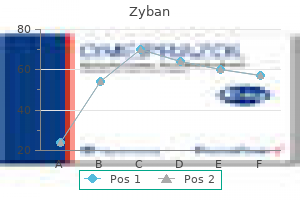
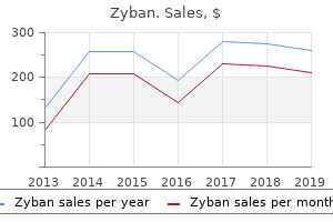
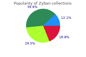
L-Carnitine Fumarate (L-Carnitine). Zyban.
Source: http://www.rxlist.com/script/main/art.asp?articlekey=96985
Protein synthesis requires that all 20 of the common 0112 amino acids be present, including the essential ones. Nutritionists evaluate the quality of a particular dietary protein by comparing its amino acid composition to that of a reference protein which has the optimum proportions of all the essential amino acids. In general, animal proteins such as beef, fish, and egg white approximate those standards. Although vegetable proteins are sometimes referred to as "incomplete" proteins, they too usually contain all the essential amino acids, but not in the ideal proportions. For example, proteins in cereals such as wheat and rice are relatively deficient in Iysine. By contrast, whereas the proteins of legumes such as peas and beans actually contain a higher level of lysine than the reference protein, they are relatively deficient in the sulfur-containing amino acids (methionine plus cysteine). However, when these two types of proteins are mixed as they would be in a meal of rice and beans, they complement each other to provide high-quality dietary protein. Improvement of the quality of vegetable proteins can also be achieved by including a small quantity of animal protein in the meal. In addition, the carbon skeletons of 11 of the other common amino acids are readily utilized for the synthesis of glucose. Instead, the pathways that catabolize their carbon skeletons generate acetyl-CoA and acetoacetate. Since hepatocytes use acetyl-CoA to synthesize ketone bodies under fasting conditions, lysine and leucine are classified as ketogenic amino acids. Five of the 20 common amino acids are both glucogenic and ketogenic, in that some of their carbon atoms can be utilized to produce glucose whereas the other carbon atoms generate ketogenic acetyl-CoA. These mixed glucogenic/ketogenic amino acids include tryptophan, isoleucine, threonine, phenylalanine, and tyrosine. Tyrosine is the precursor of the catecholamine neurotransmitters (dopamine, norepinephrine, and epinephrine), the thyroid hormones (thyroxine and triiodothyronine), and the melanin pigments. Other important amino acid-derived products include y -aminobutyric acid, histamine, nitric oxide, and polyamines. The transsulfuration pathway by which the sulfur atom of methionine is utilized to synthesize cysteine occurs primarily in liver, kidney, and intestinal mucosa. This is particularly true of neurotransmitters such as dopamine, norepinephrine, and serotonin, each of which is produced by specific groups of neurons. By contrast, the initial transamination of the branchedchain amino acids leucine, isoleucine, and valine, and the subsequent oxidation of their carbon skeletons, occur primarily in skeletal muscle. As pointed out above, the carbon skeletons of alanine and glutamine are major substrates for gluconeogenesis in liver and kidney, respectively. Glutamine is a significant fuel for rapidly dividing cells such as lymphocytes, macrophage, and enterocytes, as well as for the kidney. In the fed state, there is active protein synthesis by muscle, liver, and other tissues; excess amino acids are oxidized for energy. Most of the carbon skeletons of the branched-chain amino acids are utilized as fuel. The amino groups of the branched-chain amino acids are exported from muscle as alanine and glutamine, which are transported to the liver and kidney to provide substrates for gluconeogenesis. Rather than catalog all of these pathways, specific examples are cited to illustrate the types of reactions involved in amino acid metabolism. This process initiates amino acid catabolism by transferring the a-amino group of various amino acids to a-ketoglutarate, thereby generating glutamate. Transfer of the a-amino group of glutamate to various a-ketoacids can also be utilized to synthesize other nonessential amino acids. The precursors for this pathway are glucose, which is used to generate pyruvate, and amino acids, which donate the a-amino groups needed for glutamate synthesis. Synthesis of glutamate semialdehyde by reduction of the y-carboxyl group of glutamate to an aldehyde also serves as the initial step in the pathway of proline synthesis. The first step in the catabolism of the branched-chain amino acids is the transfer of the a-amino group of each to a-ketoglutarate,generating the corresponding branchedchain a-ketoacid. The enzymes common to catabolism of all three amino acids are 0 branched-chain amino acid transaminase and 0a-ketoacid dehydrogenase. The next step in the catabolism of the three branched-chain amino acids is an oxidative decarboxylation reaction catalyzed by the branched-chain a-ketoacid dehydrogenase complex.
The undifferentiated cells in these areas continue to divide until they become confluent, after which individual cells, the myoblasts, differentiate. The myoblasts elongate and take on the fusiform shape characteristic of adult smooth muscle cells; early in development the myoblasts resemble fibroblasts. The cytoplasm contains abundant free ribosomes, granular endoplasmic reticulum, and mitochondria, and Golgi complexes are prominent. Few filaments are present initially, but as the smooth muscle cell develops, bundles of thin filaments appear in association with dense bodies. Concomitant with the appearance of filaments, cytoplasmic organelles decrease in number. Attachment plaques along the cell membrane, plasmalemmal vesicles, and a external lamina appear at the same time or a little later than the thick filaments. In the newborn, myofilaments are small and usually restricted to the cytoplasm at the edge of the cell. Granular endoplasmic reticulum and Golgi elements are prominent and extend throughout the cell. The smooth muscle of the iris is commonly thought to be derived from cells near the margin of the optic cup and therefore is of ectodermal origin. Summary Nerve impulses reaching the myoneural junctions of skeletal muscle cells cause release of acetylcholine contained within the synaptic vesicles. This neurotransmitter diffuses across the synaptic trough, the surface area of which is greatly increased by the junctional folds. Acetylcholine causes depolarization of the sarcolemma at the motor end-plate, from which an excitation wave (action potential) sweeps over the surface of the muscle fiber and, by means of the T-system, is delivered to all the myofibrils throughout the fiber. At the triads, the impulse is passed to the sarcoplasmic reticulum, causing release of calcium ions from the terminal cisternae. Free calcium ions activate the myosin-actin interaction through the intervention of tropomyosin and troponin. Troponin acts as a calcium receptor that, in the presence of increased calcium ion, is thought to cause conformational changes in the tropomyosin, bringing about the interaction of myosin and actin. As the actin filaments slide past the myosin filaments to penetrate more deeply between them, the length of each sarcomere is reduced, resulting in an overall shortening of each myofibril and thus of each fiber. The tension generated is transmitted in succession to the sarcolemma, the external lamina, the connective tissue sheath, and then to a tendon or other muscle attachment. Muscle spindles serve as stretch receptors and coordinate the degree of muscle contraction with the strength of the stimulus. Tendon organs sense the stresses produced by the muscle contractions and prevent them from becoming excessive. The function of skeletal muscle depends on the precise alignment of actin and myosin filaments within each myofibril. A complex skeleton of accessory proteins (myomesin, -actinin, desmin, titin) supports the myofilaments, maintains their alignment, and holds them in register to each other. Intermediate filaments of the cytoskeleton (desmin) link adjacent myofibrils and maintain their register as well as linking them to the lateral aspects of the sarcolemma. Individual skeletal muscle fibers are organized and harnessed by several extracellular components (external lamina, endomysium, perimysium, and epimysium) to complete the organization of each cell into a muscle. The duration of the excitation wave at the myoneural junction is limited by the rapid breakdown of acetylcholine by the enzyme acetylcholinesterase, which is associated with the external lamina of the junctional folds. Skeletal muscle fibers have limited capacity to regenerate but can repair minor injuries. These cells, which separate from the parent muscle fiber, divide and fuse with other satellite cells to form myotubules and new fibers that bridge small defects in the parent fiber. Adult muscle increases in mass by increase in the size of existing fibers, not by increase in the number of fibers. A shift occurs from the small- and mediumsized fibers of normal muscle to the large fiber groups, but few of the hypertrophied muscle fibers become larger than the largest normal fibers. Physiologic hypertrophy at first consists of an increase in the amount of sarcoplasm followed by an increase in the number of myofibrils. Contraction of cardiac muscle occurs spontaneously, with no need for an external stimulus.
In contrast, mycotoxin levels were very low in other major corn producing states of Illinois, Iowa, Minnesota, North Dakota, and South Dakota. Some areas of Nebraska also had higher than average levels of vomitoxin in corn resulting in 1. These results indicate that corn harvested in 2011 contained primarily vomitoxin, and the majority of samples contained less than 1 ppm. Percentage of Corn Samples Containing Various Levels of Vomitoxin (Dairyland Laboratories, Inc. Percentage of Corn Samples Containing Various Levels of Aflatoxin (Dairyland Laboratories, Inc. Corn belt had growing and harvesting conditions conducive to vomitoxin production in 2011. Polioencephalomalcia is caused by necrosis of the cerebrocortical region of the brain of cattle, sheep, and goats. Hydrogen sulfide is toxic and accumulation in the rumen is thought to be the cause of these toxic effects. This causes a dramatic shift in rumen microbial populations that produce thiaminase, resulting in a thiamin deficiency. Sulfur also appears to have a significant role and interaction with thiaminase production to cause this condition, but the mechanism is not well understood. In addition, excess dietary S can interfere with copper absorption and metabolism. As a result, when high dietary levels of S are fed for an extended period of time, dietary copper levels should also be increased (Boyles, 2007). By increasing the forage content of the diet, rumen pH will not be reduced, and therefore, not favor the formation of hydrogen sulfide and allow the concentration of hydrogen sulfide to increase in the rumen. This allows nutritionists and feed formulators the ability to determine an adequate safety margin during feed formulation to manage this variability. In addition to the S content of the feedstuffs, drinking water may also be a significant source of total dietary S intake in certain geographic regions. Additional dietary S intake (%) from drinking water at various ambient temperatures and water sulfate concentrations1. Feedlot cattle appear to be most susceptible to S toxicity during the first 30 days on a finishing diet when consuming high S water or high concentrations of S in feed. This increased vulnerability to S toxicity from feeding a high concentrate, high S diet appears to be caused by a dramatic increase in rumen hydrogen sulfide concentrations which results from an increase in sulfate reducing bacteria and a decrease in rumen pH. Since sulfate-reducing bacteria utilize lactate to convert S to sulfide, the increased availability of lactate during this early finishing period may increase their metabolism and produce more hydrogen sulfide. Oxidative damage of lipids in feed negatively affect pig health and growth performance (Miller and Brzezlnska-Slebodzlnska, 1993; Pfalzgraf et al. Water sulfate content and consumption must also be considered when managing total S intake of feedlot cattle. Impact of oxidized corn oil and synthetic antioxidant on swine performance, antioxidant status of tissues, pork quality and shelf life evaluation. Effect of santoquin and oxidized fat on liver and intestinal glutathione in broilers. Feed contamination affects the entire food chain and costs millions of dollars in lost revenue and increased costs. Illness, death and potential future health risks can also occur and because we now live in a global economy, use of contaminated feed can have a global impact. Feed and food safety systems and monitoring are continually improving in many countries. This new law also prohibits shipping food or feed by interstate commerce without a current registration. Enactment of this new law will provide even greater insurance and confidence that U. While this law has not been implemented, it requires ethanol plants manufacturing corn coproducts. In general, the rule will require feed manufacturers to evaluate known or potential feed safety hazards, identify and implement preventative control procedures, monitor those procedures, take corrective actions when they are not working, and periodically verify that the overall system is working effectively. There has been a long-term scientific debate regarding the feasibility and likely efficacy of enforcing a Salmonella negative standard for animal feeds to reduce the incidence of human salmonellosis (Davies et al.
References: